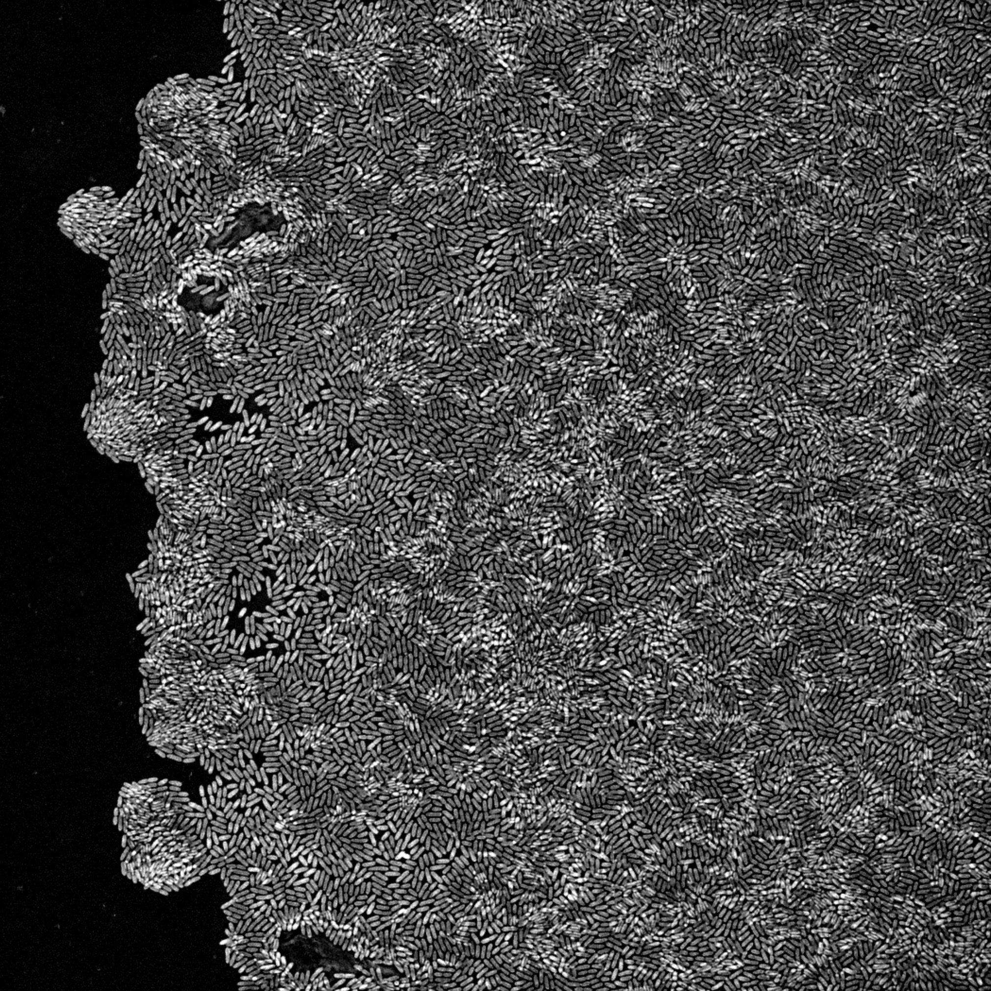
Microbial biofilms
Microbial biofilms are three-dimensional communities of bacteria and other microorganisms attached to surfaces. They are encased in a self-produced matrix of extracellular polymeric substances (EPS), representing the dominant form of microbial life in nature and the clinic. Most chronic and device-associated infections are biofilm-related.
Bacteria inside a biofilm are far harder to kill than free-floating cells. Antimicrobials diffuse slowly through the EPS matrix, and dormant persister cells can survive treatment. As a result, biofilm research has become central to modern microbiology and antimicrobial discovery.
Holotomography (HT) offers a different way to study biofilms. It is a label-free, quantitative 3D imaging technique based on refractive index, which provides natural contrast without dyes. Bacterial cells and their EPS matrix can be visualized in living samples, without staining, fixation, or photobleaching.
This makes it possible to follow biofilm formation, antimicrobial treatment, and structural changes in real time, across the full depth of the community. Time-lapse sequences can run from minutes to days. HT supports researchers in characterizing biofilm architecture and quantifying antimicrobial efficacy, including the action of antimicrobial peptides (AMPs), antibiotics, and antimicrobial nanomaterials, across both fundamental biofilm biology and applied antimicrobial development.
Features
-

- Label-free live imaging
- Visualize biofilms in their native state, without staining, fixation, or photobleaching.
-
- High-resolution 3D imaging
- Resolve EPS matrix, microcolonies, and individual bacterial cells in three dimensions, at high resolution.
-

- Long-term time-lapse imaging
- Follow biofilm formation and antimicrobial responses continuously, from minutes to days.
-
- Quantitative phenotyping
- Extract biofilm thickness, biomass, and dry mass directly from refractive-index data.
Discover Microbial biofilms with HT
-
Live observation of biofilm formation in 3D
Capturing biofilm formation live is constrained by the very imaging approaches researchers want to avoid using on early-stage cultures: staining, fixation, and dyes that perturb the community.
Holotomography removes that constraint. The label-free 3D time-lapse shown here tracks initial attachment of planktonic cells, progressive cell aggregation, and the formation of the early three-dimensional biofilm structure in Pseudomonas aeruginosa.
Data courtesy of the Yilin Wu Lab, Department of Physics, The Chinese University of Hong Kong. -
Antimicrobial peptide activity in biofilms
Evaluating antimicrobial peptide (AMP) candidates against biofilm-forming pathogens requires more than endpoint inhibition data. Researchers need a way to see, in real time, how the peptide acts on bacterial structure and on the biofilm itself.
Holotomography provides exactly that. In Kumar et al. (2025, Advanced Science), HT was used to track Hirunipin 2 against multidrug-resistant Acinetobacter baumannii biofilms. Label-free 3D imaging quantified bacterial dry mass and volume changes during peptide treatment, captured membrane disruption and intracellular aggregation leading to cell death, and measured progressive biofilm dry mass loss across the time course.
Hirunipin 2 inhibited biofilm formation and eradicated preformed biofilms (Kumar et al., 2025). -
Nanomaterial-based biofilm disruption
Evaluating antimicrobial nanomaterials against biofilms with traditional colorimetric assays is limited by metal-ion leaching that interferes with optical readouts. Investigators need a structural, label-free measurement to complement those assays.
Holotomography fills that gap. In Martínez-Montelongo et al. (2024, Ceramics International), HT combined with atomic force microscopy evaluated CuFe2O4 and CuFe2O4/ZnO magnetic nanomaterials against Candida biofilms.
Three-dimensional refractive-index imaging quantified biofilm structural disruption and intracellular damage in embedded cells (Martínez-Montelongo et al., 2024).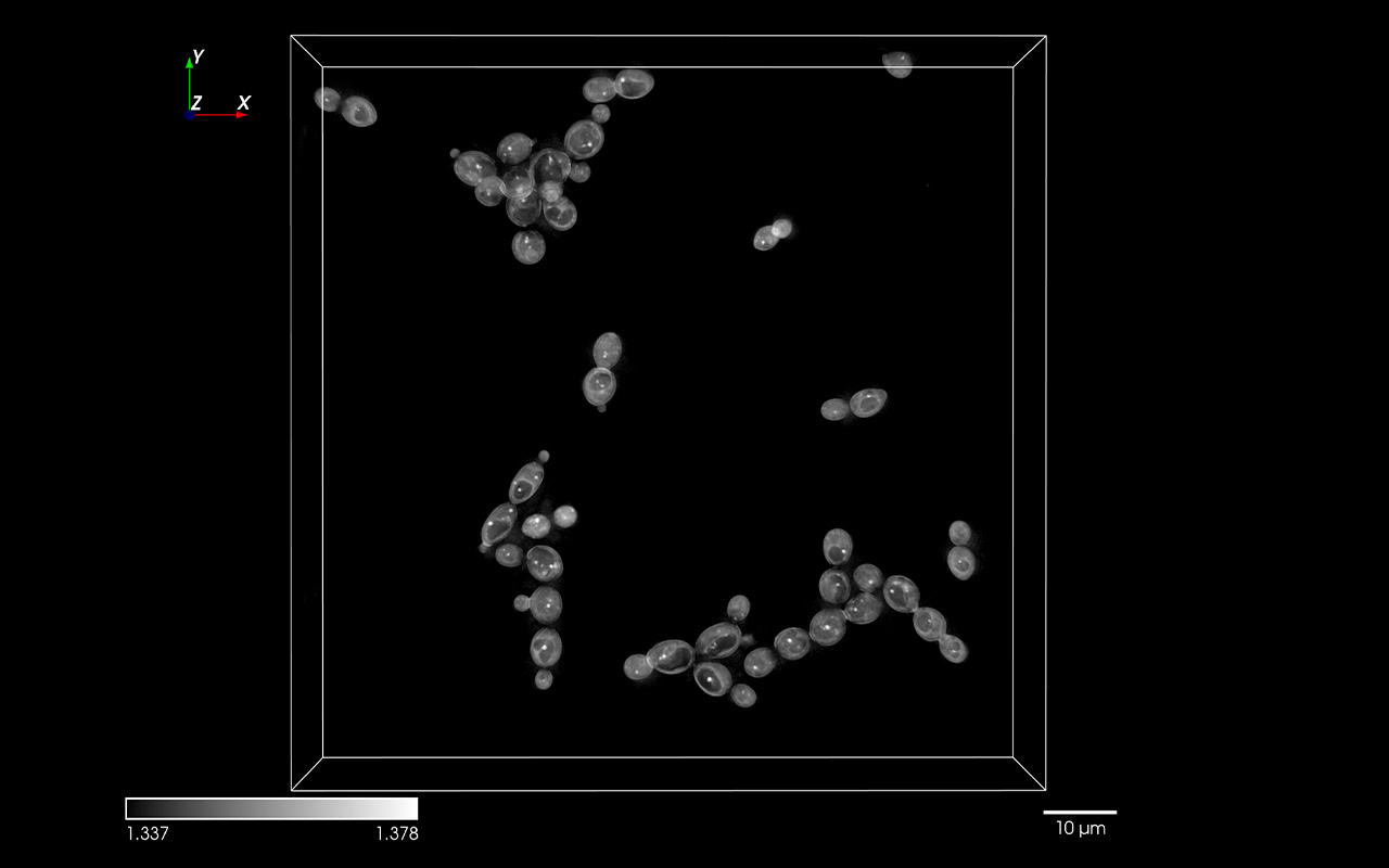
-
Biofilm formation and microbial ecology
Linking single-cell morphological adaptation to community-level functions like nitrogen fixation is difficult with bulk assays, which capture functional output but mask the cell-scale phenotypes that drive it.
Holotomography combined with fluorescence imaging bridges that gap. In Fernández-Juárez et al. (2023, Applied and Environmental Microbiology), the approach was applied to Rhodopseudomonas sp. BAL398, an anoxygenic phototrophic bacterium from the Baltic Sea.
Correlative HT–FL imaging localized nitrogenase to the center of rosette-like cellular structures and quantified a sharp increase in rosette occurrence during peak N2 fixation (Fernández-Juárez et al., 2023).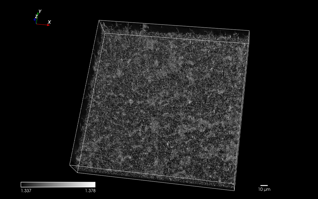
-
Beyond biofilms: bacterial intracellular phenotyping
Quantifying intracellular polymer accumulation in individual bacterial cells is a long-standing challenge in industrial microbiology. Most methods destroy the cell or rely on bulk measurements, masking strain-level differences.
Holotomography enables in vivo, single-cell quantification. In Choi et al. (2021, PNAS), HT measured polyhydroxyalkanoate (PHA) granule volume, weight, density, and intracellular localization in living cells of Cupriavidus necator and recombinant Escherichia coli.
The data revealed denser granule packing and distinct cell-pole positioning in the recombinant host versus the native producer (Choi et al., 2021).
Selected publications
-
Holotomography was used to monitor in real time how the leech-derived antimicrobial peptide Hirunipin 2 acts on multidrug-resistant Acinetobacter baumannii. Label-free 3D imaging captured bacterial membrane disruption and intracellular aggregation leading to cell death, and demonstrated that Hirunipin 2 inhibits biofilm formation and eradicates preformed biofilms.
-
Combined holotomography and atomic force microscopy were used to evaluate magnetic nanomaterial (CuFe₂O₄ and CuFe₂O₄/ZnO) activity against Candida biofilms. Three-dimensional label-free imaging resolved nanomaterial interaction with biofilm structure and intracellular damage within embedded cells, providing complementary structural readouts in cases where metal-ion release can interfere with traditional colorimetric assays.
-
Holotomography combined with fluorescence imaging was applied to study biofilm formation and morphological plasticity in Rhodopseudomonas sp. BAL398, an anoxygenic phototrophic bacterium from the Baltic Sea. Label-free 3D imaging supported correlation between cellular morphology, rosette-like structure formation, and nitrogen-fixation activity under varying light, oxygen, and nutrient conditions.
-
Label-free 3D holotomography enabled in vivo visualization and quantification of polyhydroxyalkanoate (PHA) granules in Cupriavidus necator and recombinant Escherichia coli. Refractive-index analysis provided quantitative measurements of granule volume, weight, density, and intracellular localization, revealing distinct accumulation patterns between native and recombinant PHA-producing strains.