Immunology
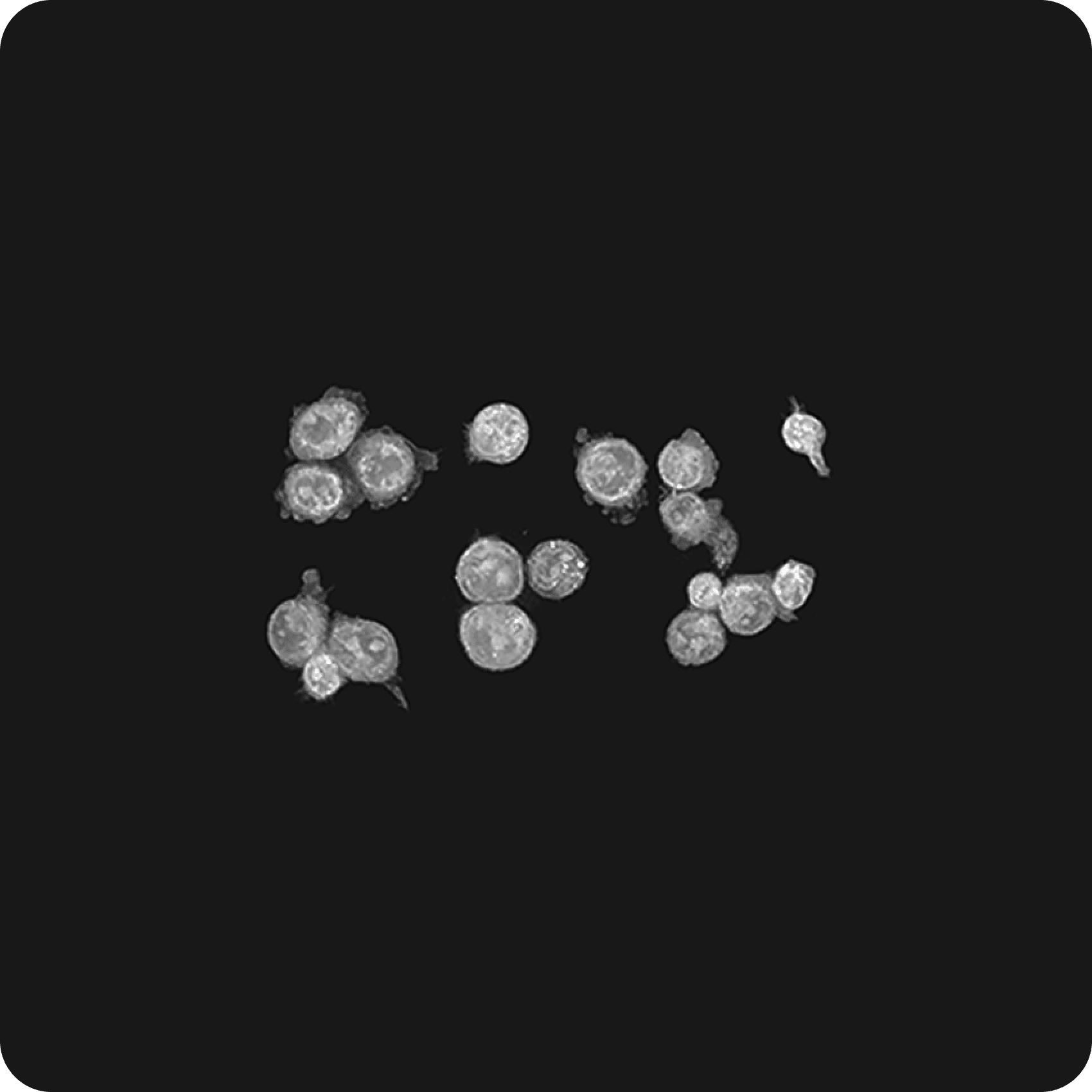
The immune response is a complex and dynamic process involving intricate interactions among various cells. To gain a comprehensive understanding of this process, it becomes crucial to observe the real time shape and movement of immune cells. In the investigation of immune responses, Holotomography (HT) emerges as a transformative tool, providing unique advantages over traditional imaging technologies.
Holotomography facilitates a detailed exploration of the immune response, allowing for the observation of immune cell shape and movement in three dimensions in real time. Its exceptional ability to instantly capture images of living cells without the need for labeling is an invaluable asset for studying immune responses. This technology also proves instrumental in deciphering processes such as how immune cells recognize and eliminate infected cells.
Features
-

- Label-free live cell imaging
- Holotomography (HT) imaging eliminates the need for labeling or dyeing, therefore minimizing artificial manipulation effects and simplifying sample preparation. This approach allows the observation of samples in their natural state, providing more reliable and relevant information.
-

- High-resolution 3D imaging
- HT provides high-content, high-resolution, three-dimensional images, allowing researchers to capture detailed information about cellular structures. This high level of detail is crucial for studying cellular morphology and dynamics.
-

- Long-term timelapse imaging
- HT facilitates continuous monitoring of cellular phenomena over extended time frames. This feature is beneficial for studying processes that unfold over hours, days, or even weeks, providing a comprehensive understanding of long-term cellular behavior.
-
- Quantitative analysis
- HT allows for quantitative analysis of cellular parameters. It can measure physical quantities such as mean refractive index,dry mass, and concentration, as well as various shape measurements like area,length, volume, and surface area.
Discover Immunology with HT
-
Immune system study
Sepsis is a severe and potentially life-threatening condition that occurs when the body's response to an infection triggers widespread inflammation throughout the body. Therefore, having a specific sepsis biomarker to enhance diagnostic accuracy and take quick action is crucial for the treatment of this condition.
In a recent study published in Light: Science & Applications, researchers suggested a promising application of 3D label-free morphology of CD8+ T cells as a potential biomarker for sepsis with remarkable accuracy and efficiency (Sung et al, 2023). When compared to currently used biomarkers, this approach offers a solution to issues like delayed responses, lack of standardization, high susceptibility, and time-intensive processes.
The proposed diagnosis and prognosis model for sepsis with a combination of Holotomography imaging for label-free 3D live cell images and deep learning advancements for engineering process simplification showcased a successful prediction rate of nearly 100%, even with only a few cells.
In another paper published in Biochemical and Biophysical Research Communications, HT was used to characterize the pathological characteristics of eosinophils in asthma patients (Kim et al., 2022). Not only subcellular organelles in eosinophils including cytoplasm, nucleus, and vacuole were well visualized without the need for labeling, immunostaining, or fixation, but specific granules were also observed and quantified to reveal insightful information into their status in asthma patients.
HT revealed that granules inside eosinophils exhibited a high molecular density characteristic based on their refractive index values. The refractive index distribution of these granules was also shown to differ between healthy individuals and asthma patients, suggesting a potential, important role of HT in clinical application for eosinophilia-associated disease by its ability to quantitively distinguish the properties of eosinophils.
Another example of HT applications in immunology involves its primary role in a 2020 study which aimed to a model for tracking and analyzing the immunological synapse (IS) dynamics of CAR-T cells (Lee et al., 2020). A combination approach of long-term, label-free, volumetric observation by HT and deep learning-based segmentation enabled an effective, automatic, and quantitative analysis of immunological synapses in 3D and in real time. The results demonstrated a new analytical model that could be generalized to study a broad range of IS for immunological research.