Regenerative medicine
Holotomography (HT) characterizes stem cells and the materials that carry them without consuming the sample, so every culture and construct can be reused after measurement.
In regenerative medicine, the cells being studied are also the cells that will be used. Most characterization assays, from immunostaining to RNA sequencing, require the sample to be fixed or lysed, so quality control usually relies on a separate culture that is sacrificed for the measurement.
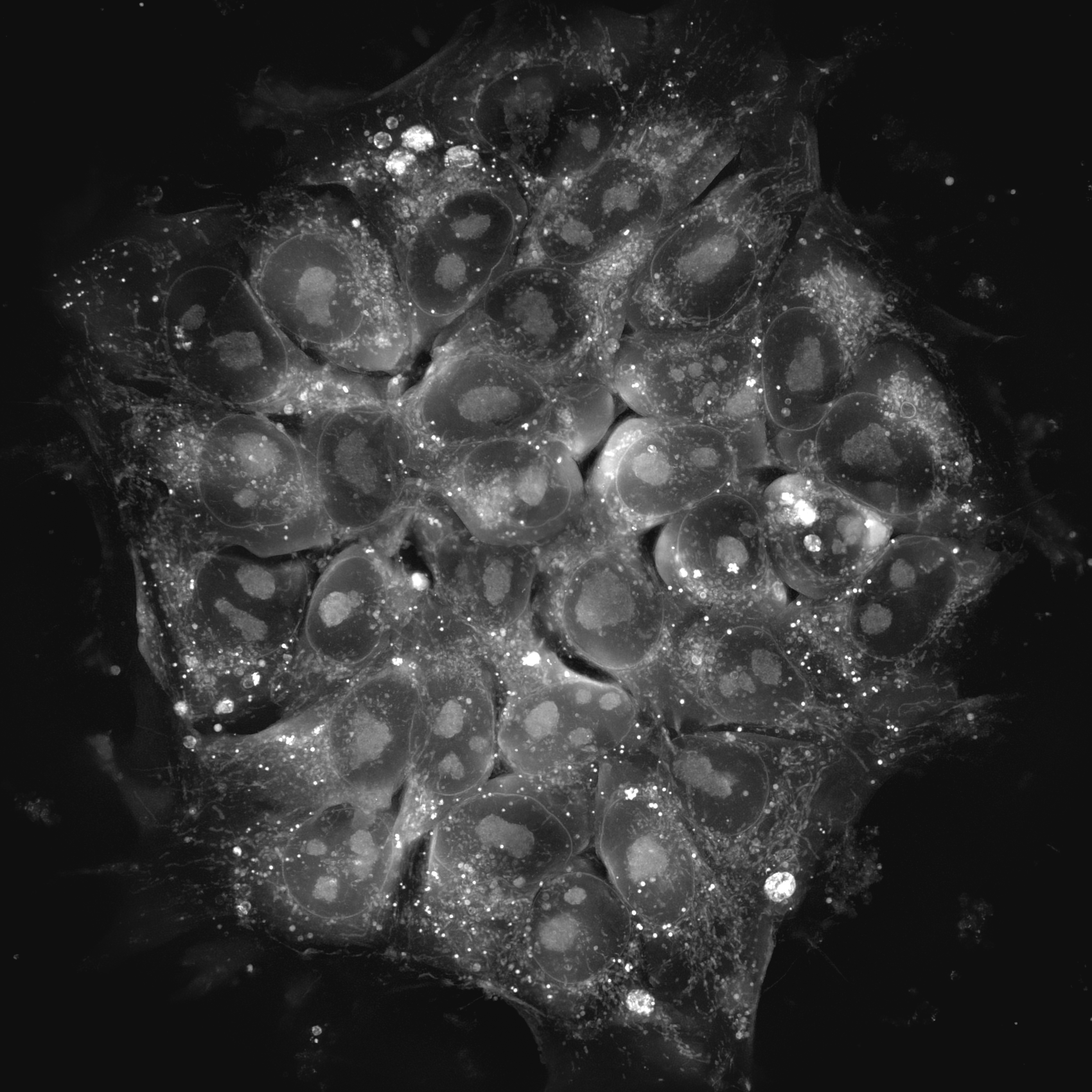
HT measures the culture that will be used, rather than a sacrificial replicate. A form of quantitative phase imaging, it reconstructs a three-dimensional refractive index tomogram of living cells and transparent materials with no exogenous dye, fixation, or fluorescent reporter line. A human pluripotent stem cell (hPSC) colony screened on day 3 can be passaged, differentiated, and imaged again on day 30.
Refractive index is proportional to local biomolecular density, so each tomogram is also a quantitative record. The same image shows how a colony flattens as it exits pluripotency and how lipid droplets and mitochondria change during differentiation. In hydrogels, it resolves particle distribution and layer structure through the full thickness without sectioning.
Results therefore arrive during the protocol, not after it. A differentiation that is drifting or a construct with a structural defect can be corrected while the sample is still in use.
Features
-

- Label-free live cell imaging
- Holotomography (HT) imaging eliminates the need for labeling or dyeing, therefore minimizing artificial manipulation effects and simplifying sample preparation. This approach allows the observation of samples in their natural state, providing more reliable and relevant information.
-
- High-resolution 3D imaging
- HT provides high-content, high-resolution, three-dimensional images, allowing researchers to capture detailed information about cellular structures. This high level of detail is crucial for studying cellular morphology and dynamics.
-

- Long-term timelapse imaging
- HT facilitates continuous monitoring of cellular phenomena over extended time frames. This feature is beneficial for studying processes that unfold over hours, days, or even weeks, providing a comprehensive understanding of long-term cellular behavior.
-
- Quantitative analysis
- HT allows for quantitative analysis of cellular parameters. It can measure physical quantities such as mean refractive index,dry mass, and concentration, as well as various shape measurements like area, length, volume, and surface area.
Discover Regenerative medicine with HT
-
AI-assisted quality control for pluripotent stem cells
Pluripotency is normally confirmed by molecular assays that destroy the culture, or by fluorescent reporters that require fixation or phototoxic exposure. Neither can monitor a colony continuously.
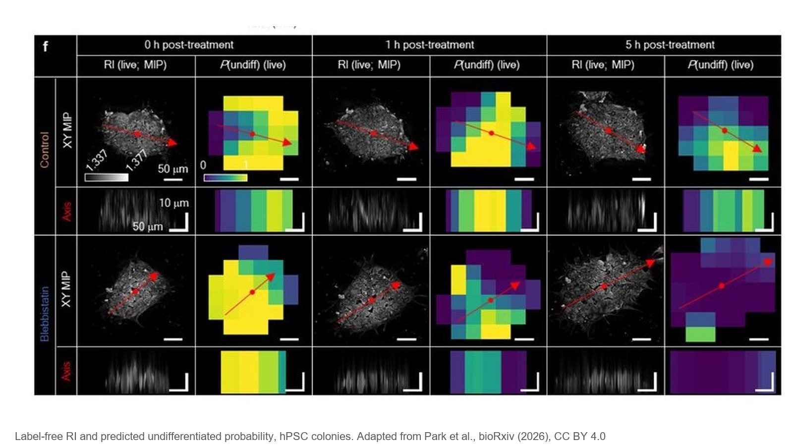
Holotomography removes that constraint. A neural network trained on 3D refractive index tomograms, each acquired in 1 to 3 minutes, detected colony flattening 6 to 12 hours before molecular markers declined, with 96.8% accuracy across multiple hPSC lines.
The screened colony stayed in culture and remained available for use (Park et al., 2026, preprint).
-
Tracking hepatic differentiation of mesenchymal stem cells
A differentiation protocol can activate the correct genes and still fail, because the block sits at the organelle level that transcriptomic snapshots do not reach.
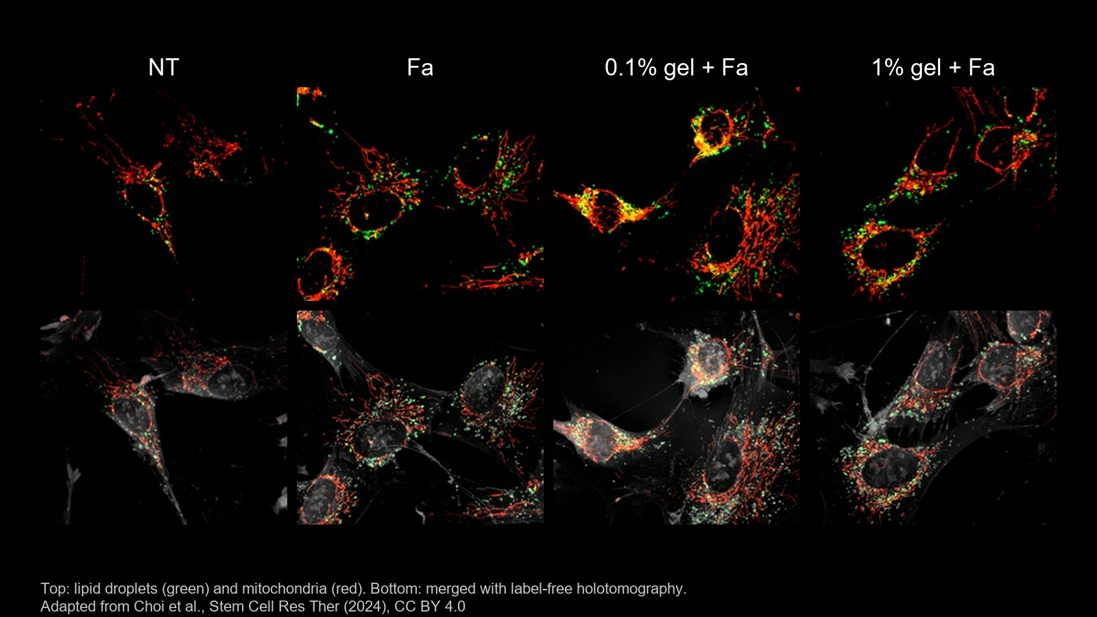
Holotomography resolves that level directly. The ROCK inhibitor fasudil switched on endoderm genes in umbilical cord mesenchymal stem cells but stalled hepatic differentiation, and HT identified the cause as excessive lipid droplet accumulation impairing mitochondrial function.
High-viscosity gelatin coating suppressed the droplets and restored mitochondrial activity, while the opposite viscosity proved better for final maturation (Choi et al., 2024).
-
Stem cell migration on an engineered culture surface
A xenogeneic-free surface has to be shown to support active migration, not only attachment, and fluorescent labels can alter the motility being assessed.
Holotomography measures it without labels. Intestinal stem cell colonies on a new xenogeneic-free polymer surface were imaged every 2.5 minutes for 6 hours, tracking colony centroids and resolving lamellipodia directly.
Colonies on the engineered surface migrated 1.8-fold faster and formed lamellipodia; colonies on untreated surfaces formed none (Park et al., 2025).
-
Non-destructive characterization of an oxygen-releasing hydrogel implant
Verifying that a therapeutic hydrogel is built as designed normally means sectioning it, which destroys the construct intended for use.
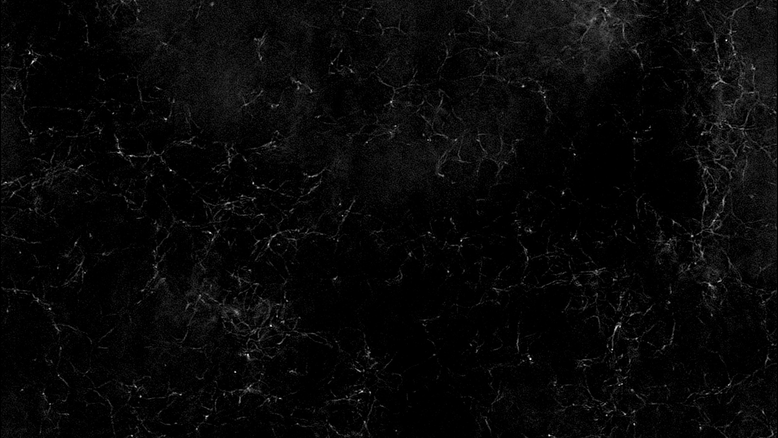
Holotomography inspects it intact. Refractive index imaging optically sectioned a full 200 µm oxygen-releasing gel, confirming uniform distribution of the oxygen-generating microparticles and resolving a core-shell microstructure by refractive index difference alone, 1.352 in the shell against 1.343 in the core.
The verified implant improved flap survival, blood flow, and mitochondrial biogenesis in a large-animal model (Jeon et al., 2025).
-
Linking biomaterial stiffness to stem cell fate
Substrate stiffness can direct stem cell fate with no biochemical cue, but demonstrating it requires quantifying intracellular change in single cells.
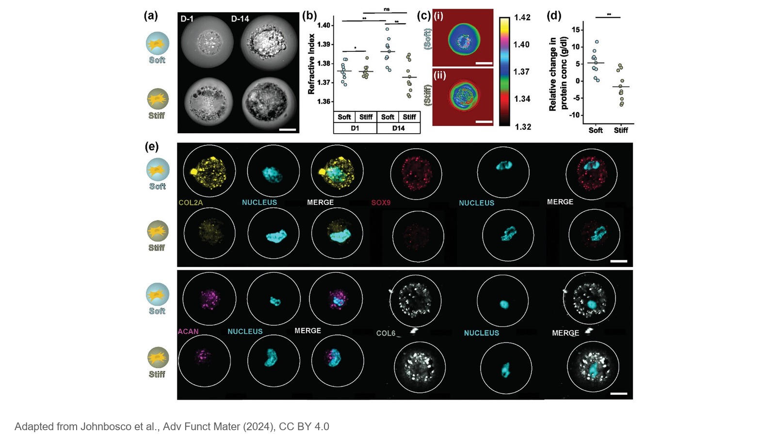
Holotomography provides that measurement. Individual mesenchymal stem cells were covalently tethered inside soft or stiff hydrogel microgels, isolating stiffness as the only variable, and refractive index was converted into protein density and dry mass.
After two weeks of chondrogenic differentiation, cells in soft microgels had deposited more nascent protein, and cells in stiff microgels showed higher molecular crowding (Johnbosco et al., 2024).
-
Mitochondrial morphology in aged bone repair
Mitochondrial dysfunction is a leading suspect in age-related loss of bone repair, and the dyes normally used to image mitochondria add phototoxic stress to cells already under metabolic strain.
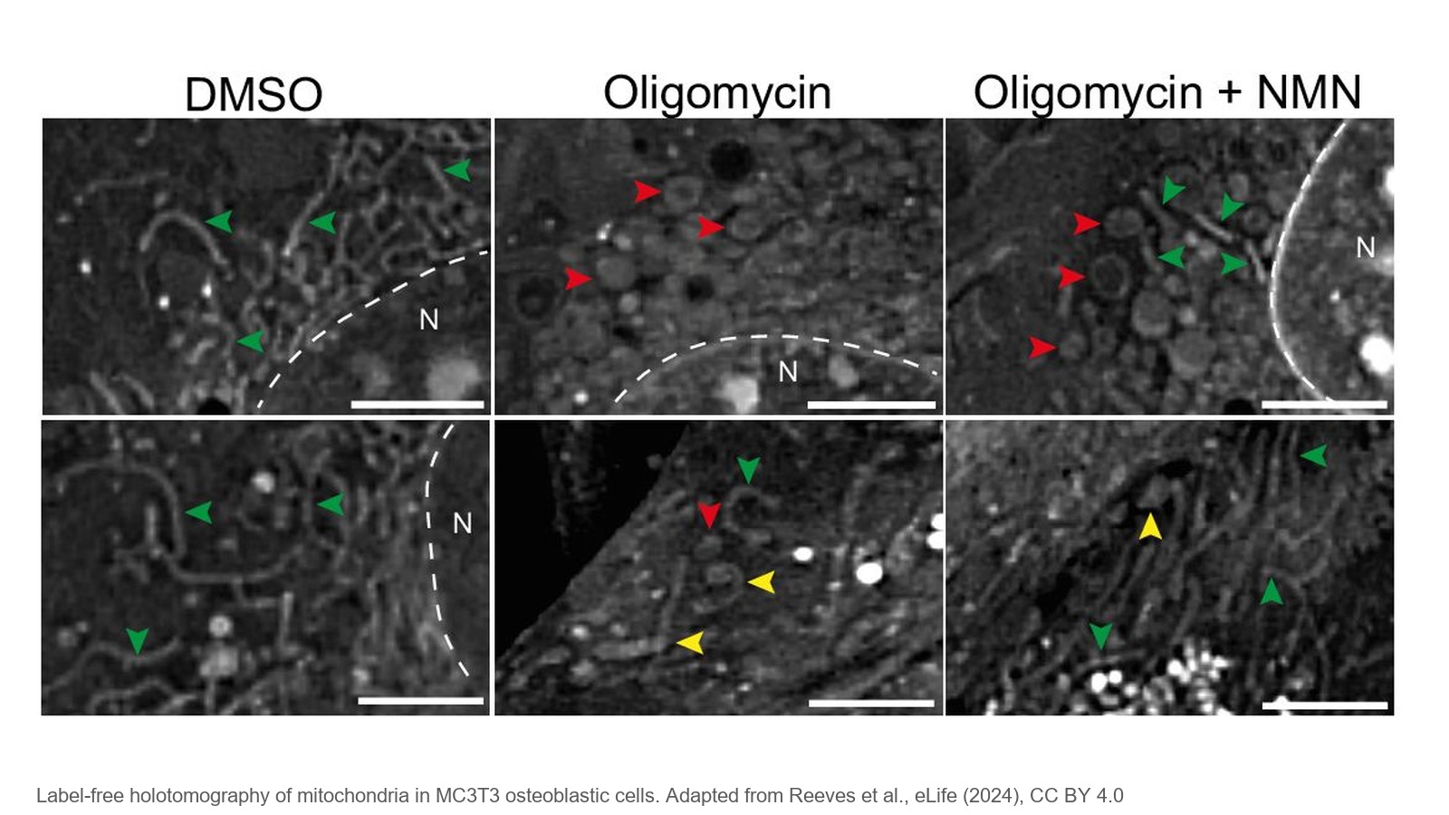
Holotomography images them without dyes. Mitochondrial morphology in osteoblastic cells was classified as round, intermediate, or rod-like and cross-validated against MitoTracker. Oligomycin shifted mitochondria toward rounded, dysfunctional forms, while nicotinamide mononucleotide (NMN) preserved rod-like morphology and ATP output.
NMN went on to restore bone repair in aged mice (Reeves et al., 2024).