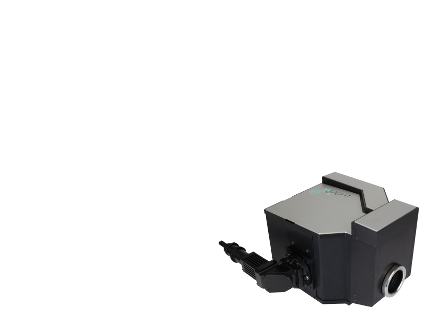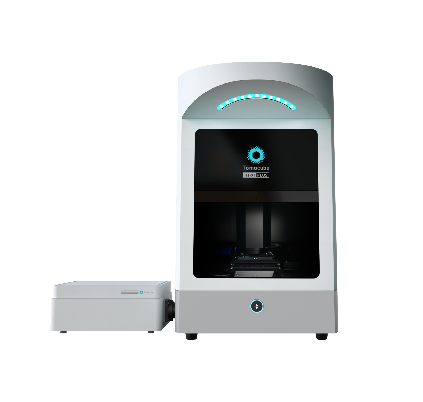
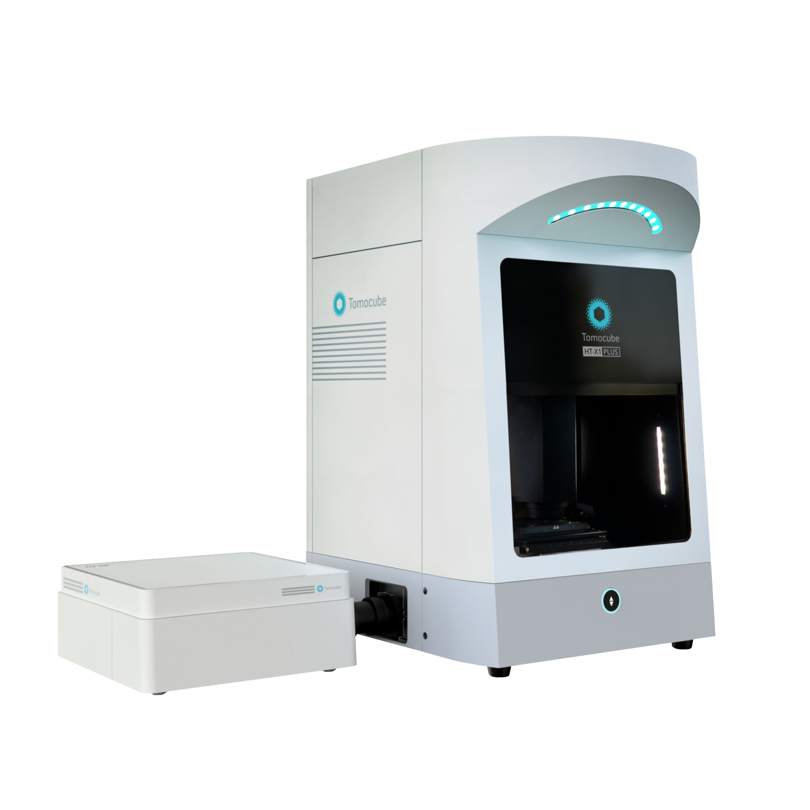
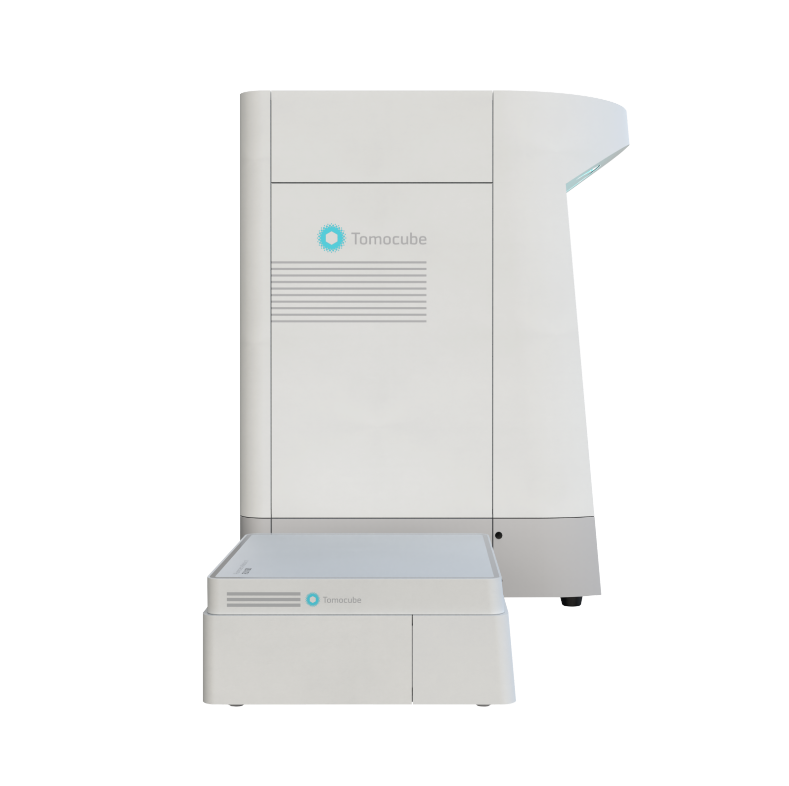
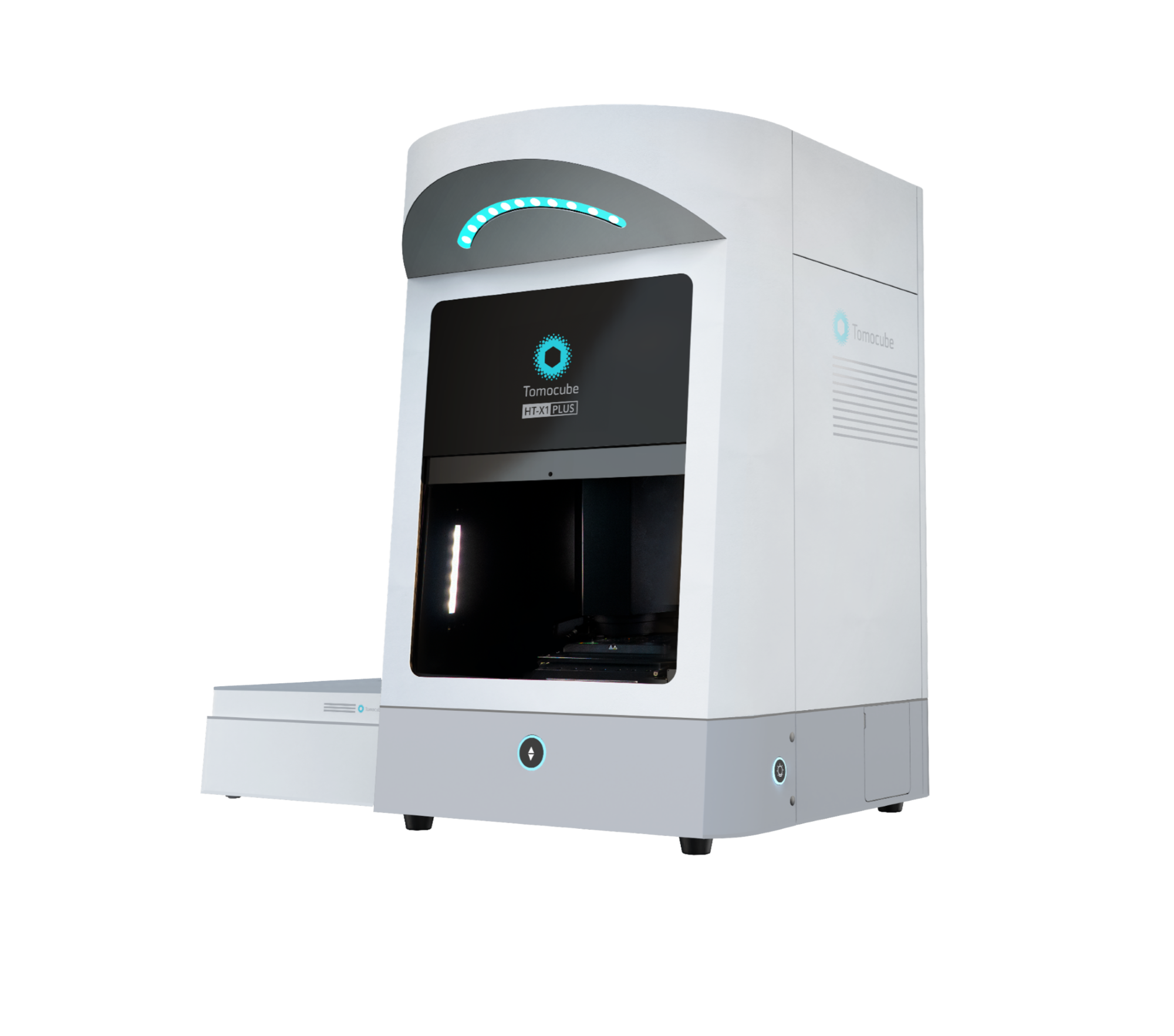
HT-X1™ Plus
Experience Exceptional Quality with our High-Performance HT Platform.
Elevate the Possibilities with Enhanced Capabilities.
The HT-X1™ Plus takes bioimaging to the next level, building on the proven success of the HT-X1™.
As the first of its kind in the 2nd-generation Holotomography series, the HT-X1™ revolutionized biomedical research with its high-resolution, 3D imaging and unparalleled stability. Its versatile platform, compatible with various imaging plates and powered by the advanced TomoAnalysis™ software, provided researchers with powerful tools for detailed quantitative analysis and reliable imaging across a wide range of applications.
Now, the HT-X1™ Plus propels this pioneering technology even further, offering enhancements designed to meet the ever-evolving demands of biomedical research. Experience clearer, more detailed imaging of multi-layered specimens with improved illumination optics and advanced image reconstruction algorithms.
Equipped with a high-spec camera featuring a 4x larger field of view and significantly reduced acquisition time, the HT-X1™ Plus is perfect for high-throughput phenotypic screening of cells and organoids. Its upgraded correlative imaging capabilities—incorporating an sCMOS-based fluorescence module—enable seamless integration of molecular studies with single-cell-resolution 3D images.
The HT-X1™ Plus extends the reach of Holotomography to an even broader array of challenging specimens, including dense organoids, tissue sections, and fast-moving microorganisms. It is a state-of-the-art Holotomography imaging platform, designed to empower researchers with the precision, efficiency, and reliability needed to drive the future of biological and biomedical discovery.
Features
-
- Large field-of-view
- Capture expansive areas without the need for stitching, ideal for large-scale, high-content experiments.
-

- Faster image acquisition
- Efficiently acquire high-content images, making it well-suited for high-throughput screening and monitoring dynamic specimens.
-
- Flexible choice of light source
- Customize your imaging with three wavelength options to enhance contrast or improve penetration, tailored to your research needs.
-

- Combine advanced fluorescence
- Maximize 3D imaging quality by integrating HT with the sCMOS-equipped fluorescence module, delivering cutting-edge 3D fluorescence imaging.
-
- Wide preview + Color brightfield
- Gain deeper insights into tissue section studies with wide preview scan mode paired with correlative color brightfield imaging
User benefits
-
High Content Screening of Live Cells and Organoids
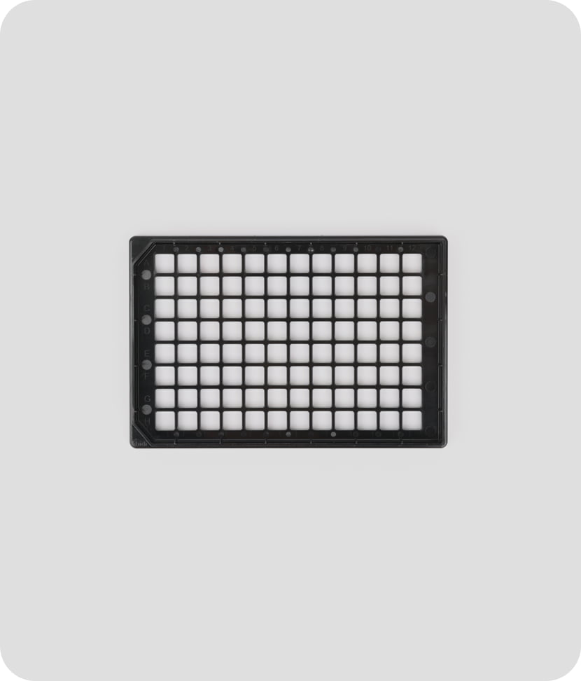
The HT-X1™ Plus is optimized for high-throughput screening, making it highly suitable for high-content, image-based drug screening research. Featuring a high-performance CXP camera and AI-powered image reconstruction algorithms, the platform excels in both coverage and acquisition speed. Its large field of view measuring 308 μm x 308 μm and rapid 3D scanning capability allows researchers to efficiently analyze an entire 96-well plate in under 30 minutes. This efficiency enables large-scale experiments with unmatched precision and consistency, resulting in faster, more reliable data acquisition that significantly accelerates the drug discovery process.
-
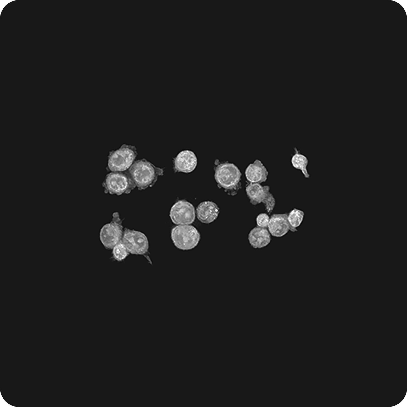
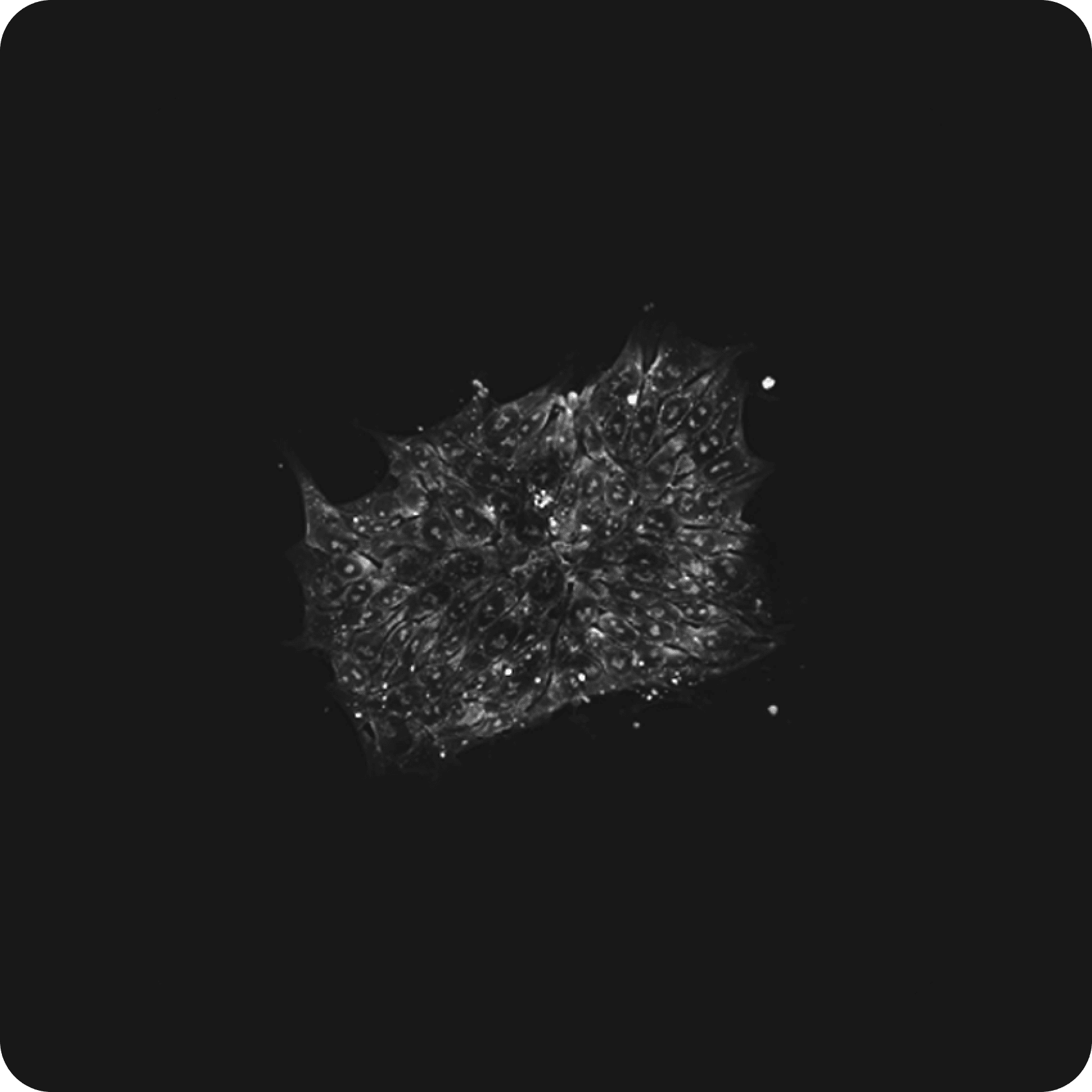
High Resolution Imaging of 3D Biological Samples
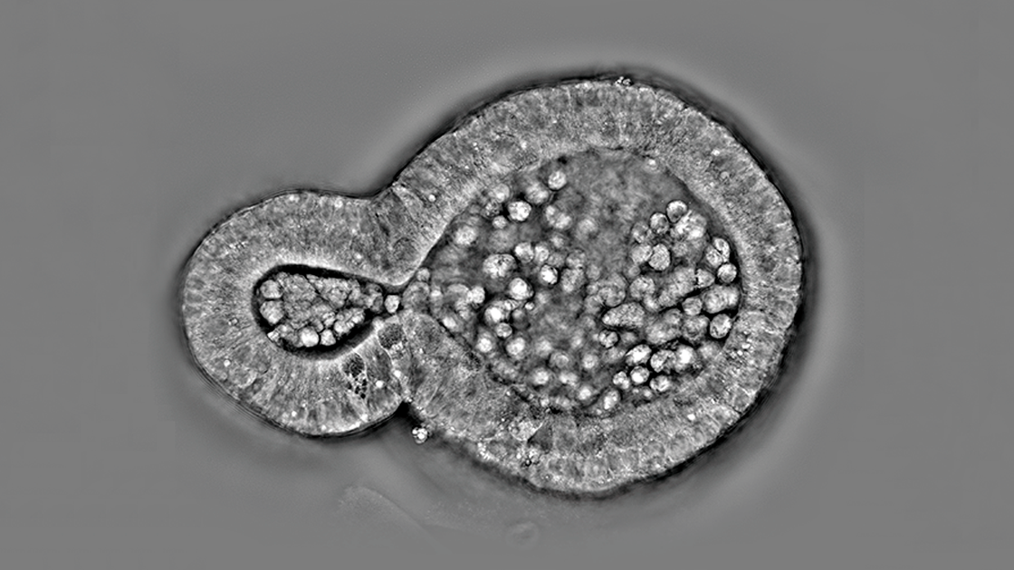
The new features of the HT-X1™ Plus are especially advantageous for research involving 3D cultures, enabling the detailed investigation of dense organoids and intact tissue sections. The platform incorporates long-wavelength light sources, improving penetration depth and reducing scattering noise, to achieve clearer 3D visualization. Additionally, an advanced 3D reconstruction algorithm further enhances both imaging clarity and precision, delivering superior image quality for 3D biological imaging.
-
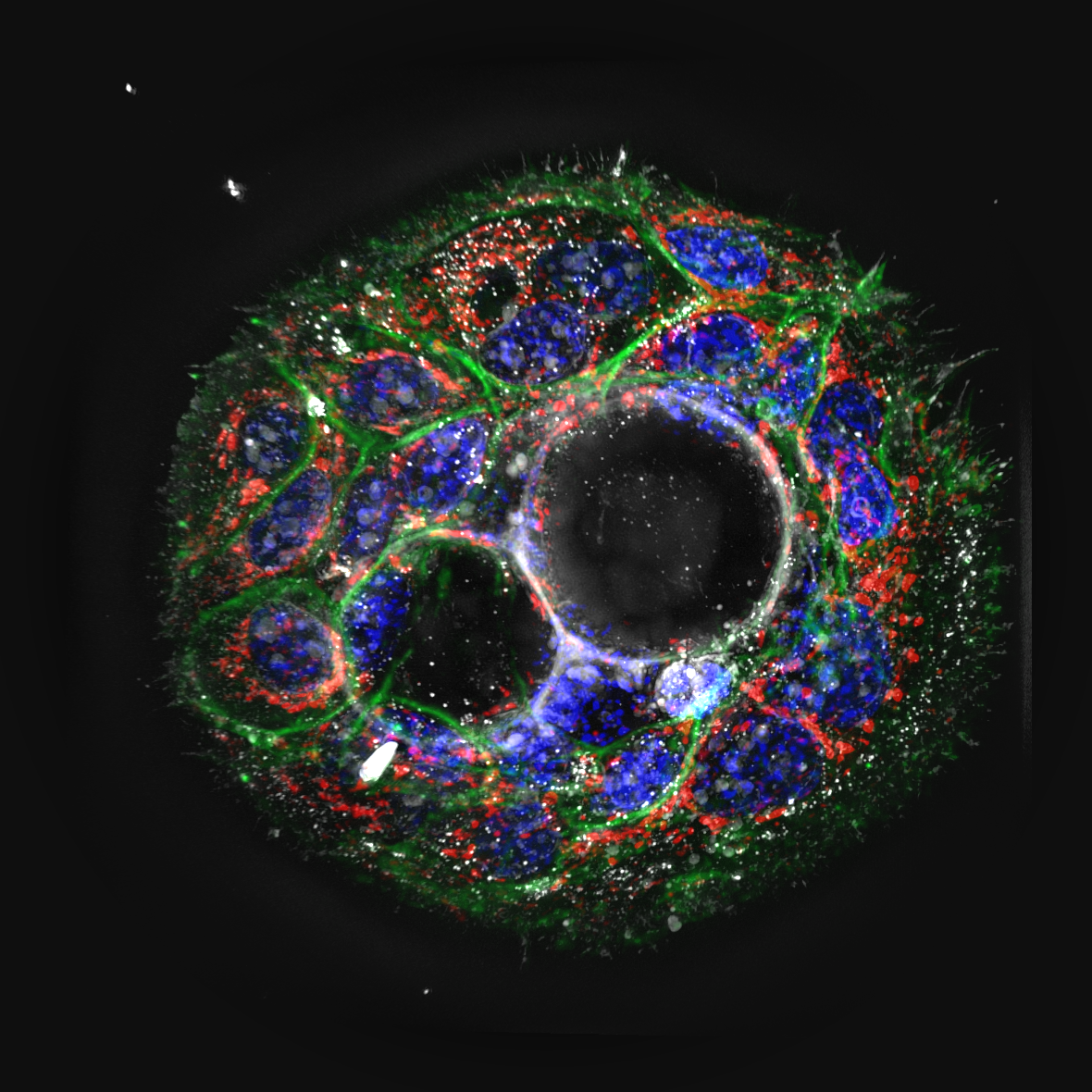
Enhanced Correlative Fluorescence Imaging
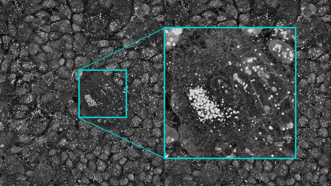
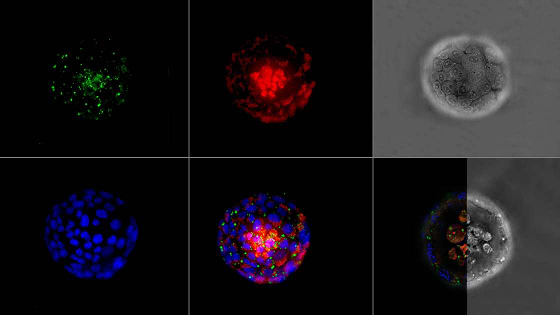
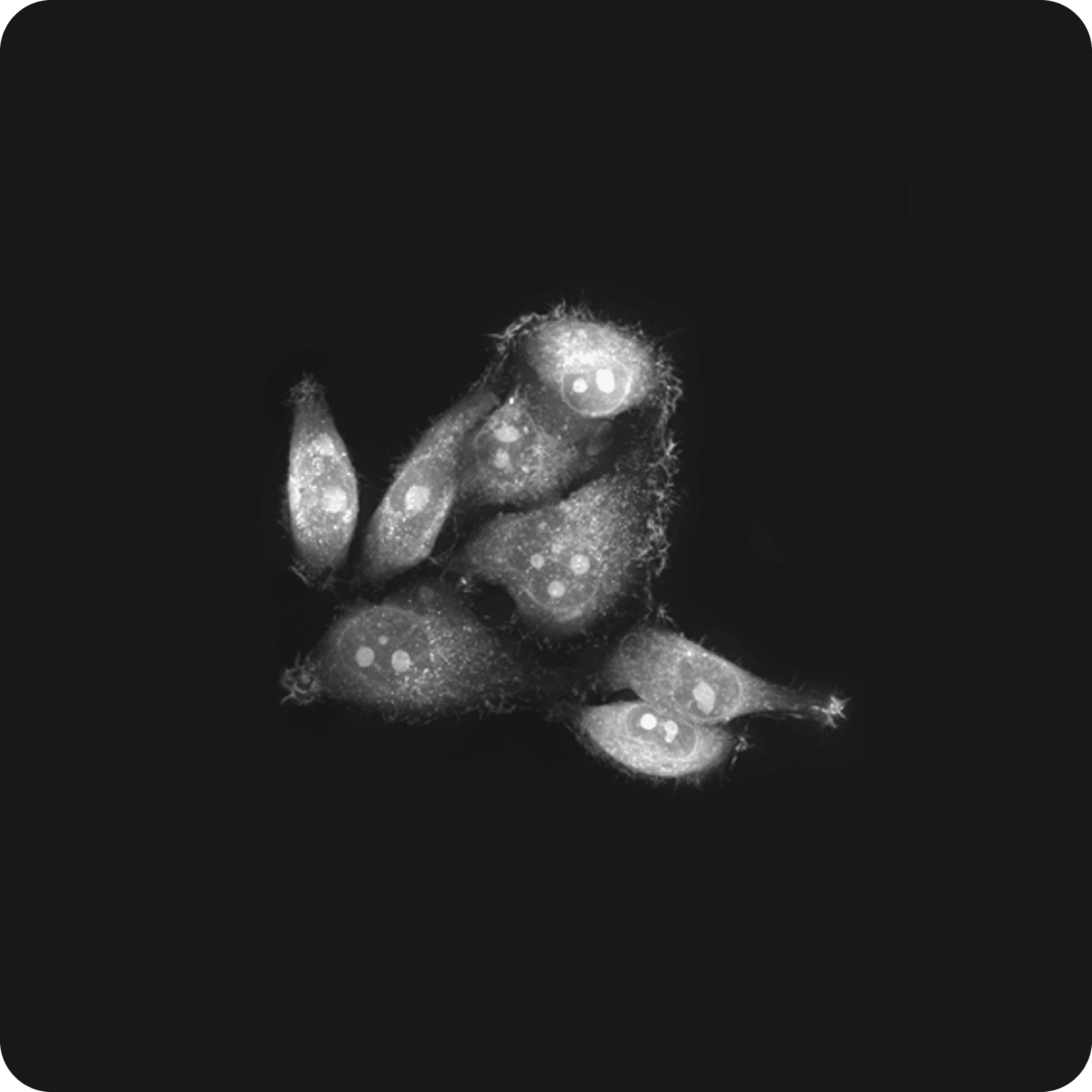
The HT-X1™ Plus offers enhanced multimodal imaging capability with its fluorescence module (FLX™) featuring an sCMOS camera designed specifically for precise signal intensity measurements. The FLX™ module offers high sensitivity to fluorescence, achieving better signal-to-noise ratio (SNR) and shorter exposure times. This allows researchers to obtain biomolecular specificity information from target organelles or fluorescence sensors, even in samples with weak fluorescent signals, such as antibody reactions or hard-to-stain organoids.
-
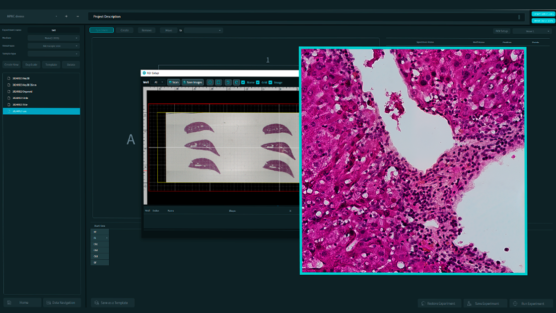
Color Brightfield Imaging for Histological Studies
With the new color brightfield imaging modality and wide preview scan features, researchers can gain deeper insights into tissue section studies. The platform allows for the seamless integration of complex structural data, obtained through 3D optical sectioning, with rich histological information from H&E staining or immunohistochemistry. This integration enhances our understanding of tissue morphology and dynamics, accelerates advancements in clinical pathology and diagnostics, and helps pave the way for the future of personalized medicine.
Applications
Configuration
-
HT-X1 Plus main unit
- Objective Lens 40× NA 0.95 Air
- Image Sensor 20 MP CMOS (CXP)
- Illumination LED 444/520/660 nm
- Motorized Stage
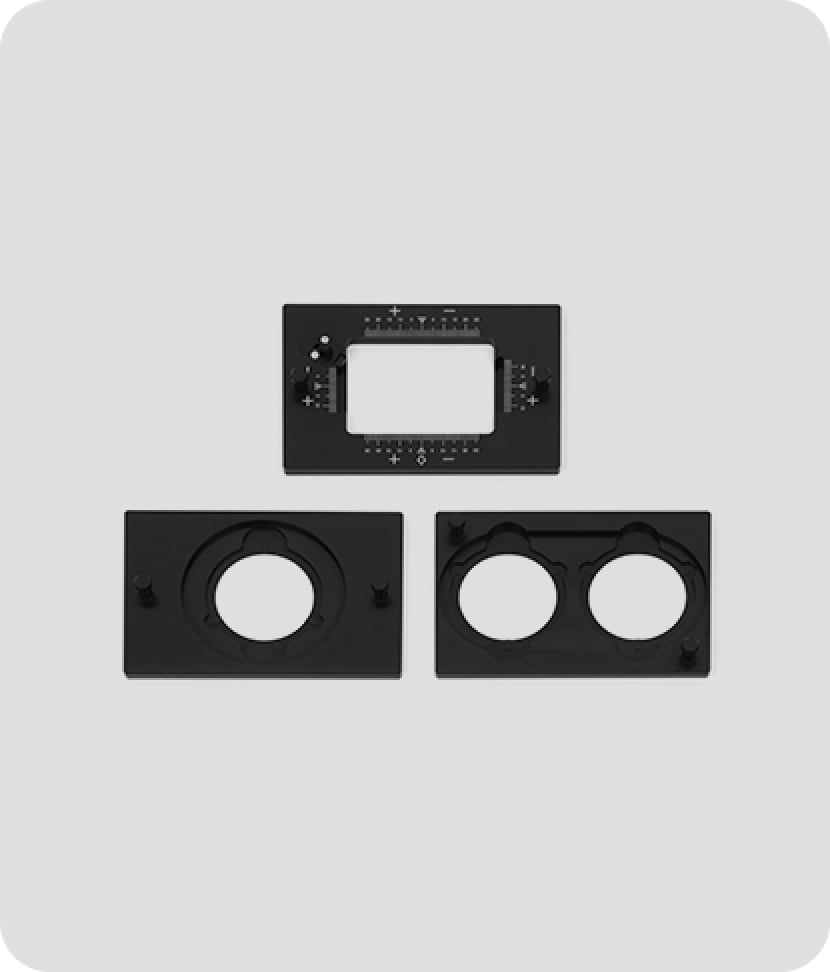
- Fluorescence Module External module equipped
- Laser-based consistent focus
- Environmental Controller

-
Fluorescence Module X (FLX 100)
- High-sensitivity external fluorescence module
- Light Source Multi-LED (DAPI/FITC/TRITC/CY5)
- Image Sensor 4.2 MP sCMOS (QE 95%)
- 4 excitation / 4 emission filters
-
X-Light V2 Spinning Disk Confocal (SDC)
- External widefield and confocal module
- Light Source 4-line lasers (DAPI/FITC/TRITC/CY5)
- Image Sensor 4.2 MP sCMOS (QE 95%)
- 8-position emission filter wheel