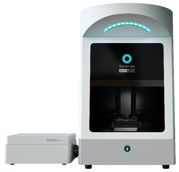
Transforming 3D Biology Using AI: Tomocube’s HT-X1™ Plus Accelerates Cellular and Organoids Label-Free Analysis
Tomocube, a global leader in 3D label-free imaging and analysis, proudly introduces the HT-X1™ Plus, a state-of-the-art bioimaging platform designed to meet the evolving needs of biomedical researchers. This new system raises the bar in high-resolution, high-throughput 3D imaging for cells and organoids, providing researchers with faster, more detailed, and more accurate insights into biological processes.
With the HT-X1™ Plus, Tomocube has integrated cutting-edge technologies that enhance precision, imaging speed, and data richness. The platform offers a 4x larger field of view and advanced illumination optics, making it a game-changer for phenotypic screening, tissue section imaging, and the study of fast-moving microorganisms. These capabilities unlock new research possibilities in cell biology, regenerative medicine, and organoids research.

Setting New Standards in 3D Imaging
Building upon the proven success of the HT-X1™, the HT-X1™ Plus sets new standards in 3D imaging performance. Equipped with a high-performance CXP camera and AI-powered image reconstruction algorithms, the platform drastically reduces scan times, enabling comprehensive 3D imaging of a full 96-well plate in under 30 minutes—without sacrificing detail or quality. “With the HT-X1™ Plus, researchers can now explore complex biological samples like organoids with unparalleled clarity and speed,” said YongKeun (Paul) Park, Chief Executive Officer in Tomocube. “This platform is designed for high-content, image-based drug screening assays and holds tremendous potential to accelerate discoveries in biomedical research.”
Key Features of the HT-X1™ Plus Include:
- Larger Field-of-View: Capture expansive areas without stitching, ideal for large-scale, high-content experiments.
- Faster Image Acquisition: Optimized for high-throughput screening, capable of scanning 96-well plates in just 30 minutes.
- Flexible Light Source Options: Configure imaging with three wavelengths (R/G/B) for optimal contrast and penetration.
- Advanced Correlative Fluorescence Imaging: Integrated sCMOS-based fluorescence module (FLX™) enhances 3D imaging with high sensitivity and precision.
- Color Brightfield Imaging: A new modality with wide preview scan mode improves histological studies, providing comprehensive tissue morphology analysis.
Solving Major Research Challenges:
The HT-X1™ Plus directly addresses key challenges in bioimaging, including slow scan speeds, limited fluorescence sensitivity, and challenges related to light absorption in thick samples. Its advanced imaging capabilities—such as reduced phototoxicity and improved 3D optical sectioning—make it the ideal tool for investigating dynamic, sensitive samples without compromising image quality.
Pushing the Boundaries of Biomedical Discovery:
Designed to integrate effortlessly with molecular studies and fluorescence-tagged sensors, the HT-X1™ Plus enables researchers to visualize and quantify intricate biological processes with unprecedented detail. The platform’s AI-driven TomoAnalysis™ software facilitates in-depth analysis, further pushing the boundaries of cellular dynamics research.
Applications of HT-X1™ Plus Include:
- Cell Biology & Regenerative Medicine: Perform high-content screening of live cells and organoids for drug discovery and therapeutic studies.
- 3D Biology & Organoids: Obtain detailed imaging of dense organoids, tissue sections, and immune cell interactions.